

EFFICIENT | INFORMATIVE | EASY
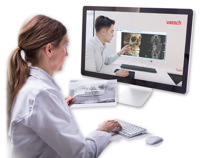
Book a VIRTUAL DEMO with your
Vatech Sales Representative Today!

Save time by scheduling a virtual demo at home or in your office.

Our intuitive user-friendly devices make it easy to learn
without in-person assistance.
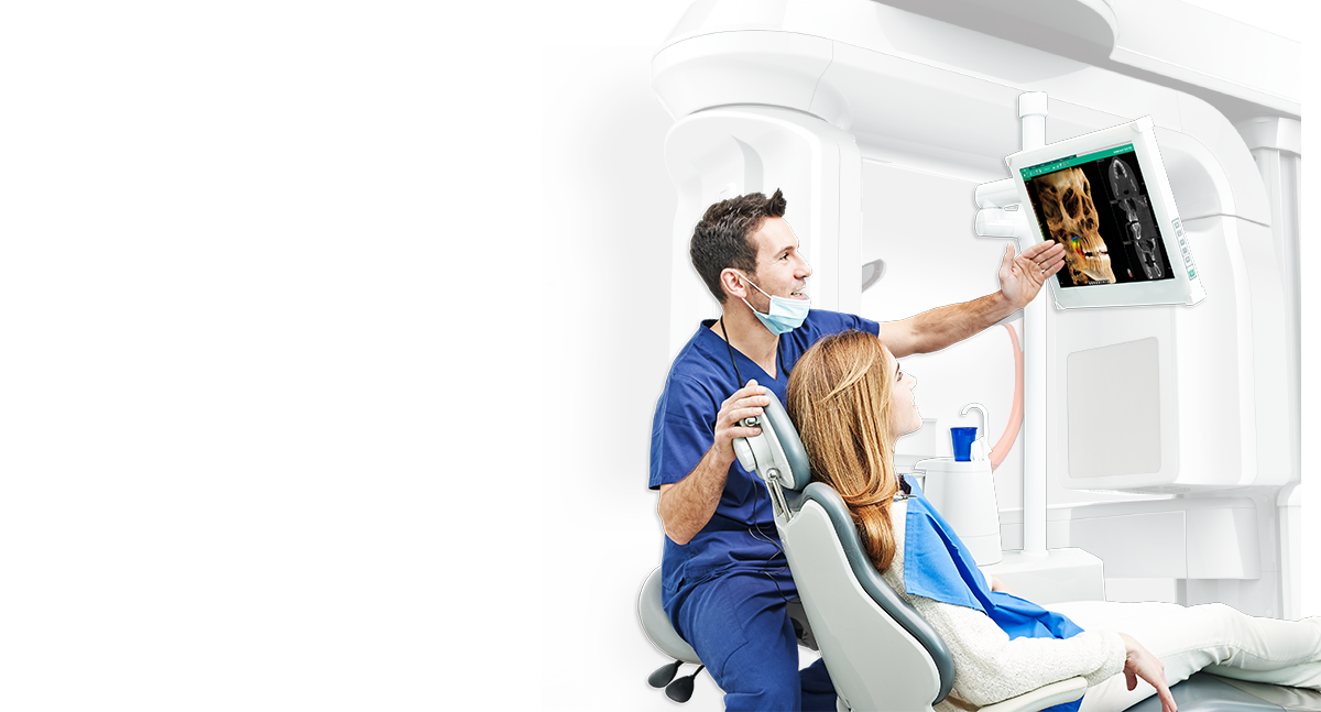
As the US subsidiary of Vatech, Inc, Vatech America is committed to providing the industry with innovative dental x-ray
imaging solutions while maintaining a primary focus on ultimately enhancing the quality of patient care.


We have numerous sales teams throughout the United States of America.
Find a Vatech Representative Near YouGet help from our technical support professionals. We are here to help.
Remote Assistant